Role of nephrologists in Diagnostic Angiography and Angioplasty
Prof. (Dr.) Debabrata Mukherjee | Renal Vascular Interventions
Senior Director – Nephrology & Renal Transplant, Medanta – The Medicity, Gurugram
Advanced Renovascular Care: Correcting Renal Stenosis
When severe arterial blockages limit critical blood delivery to the kidneys, it triggers a specialized pathological state known as **Renovascular Hypertension** and progressive nephron loss. Conditions such as **Atherosclerotic Renal Artery Stenosis (ARAS)** and **Fibromuscular Dysplasia (FMD)** restrict perfusion, signaling the kidneys to release excessive renin, which sharply raises systemic blood pressure.
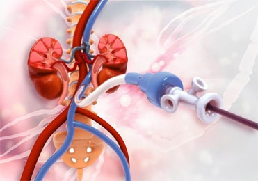
Managing these vascular conditions requires expert diagnostic **Renal Angiography**. If a significant blockage is confirmed, a micro-catheter is utilized to perform a **Renal Angioplasty with Balloon Expansion and Stenting**. Under the clinical supervision of **Prof. (Dr.) Debabrata Mukherjee at Medanta Gurugram**, these targeted endovascular procedures are executed to safely re-establish blood flow, protect kidney tissue, and stabilize severe hypertension.
Clinical Indications for Renal Angiography
Vascular mapping and interventional planning are highly recommended for patients presenting with specific clinical conditions:
- Severe, resistant hypertension uncontrolled by 3 or more medications.
- Sudden, unexplained deterioration of eGFR after initiating ACE inhibitors or ARBs.
- Significant size asymmetry (Atrophy) between the two kidneys on ultrasound.
- Recurrent, unexplained flash pulmonary edema (Bilateral Stenosis signature).
- Progressive ischemic nephropathy in patients with known peripheral vascular disease.
The Nephrologist’s Clinical Protocol at Medanta
Identifying high-yield vascular lesions through non-invasive Doppler ultrasound or CT angiography to confirm clear therapeutic benefits.
Designing personalized intravenous hydration regimens to protect existing nephrons from contrast-induced toxicity.
Coordinating directly with interventional specialists inside the cath lab to manage hemodynamic fluctuations and pressure gradients.
Monitoring post-stenting eGFR improvements and adjusting antiplatelet and antihypertensive drug combinations systematically.
Operative Logistics & Risk Defenses
The procedure involves gaining entry through the radial or femoral artery under local anesthesia. A fine guide-wire is directed into the narrow segment of the renal artery under fluoroscopic control. A high-pressure balloon is inflated to dilate the lumen, followed by the deployment of a permanent metallic stent to maintain long-term vessel openness.
Complication Control Framework:
- Contrast-Induced AKI (CI-AKI): Controlled by managing hydration volumes and using low-osmolar dye alternatives.
- Atheroembolic Micro-Lesions: Prevented by using gentle catheter manipulation techniques to minimize cholesterol plaque disruption.
- In-Stent Restenosis: Managed using periodic duplex ultrasound scanning at our Gurugram center.
Expected Clinical Benefits of Revascularization
- Significant drops in systemic blood pressure, reducing reliance on multiple high-dose medications.
- Long-term stabilization of filtration function, effectively delaying the progression toward dialysis.
- Sustained improvement in myocardial perfusion, lowering cardiorenal and flash edema risks.
- Restoration of healthy blood flow symmetry across the left and right kidney systems.
Clinical FAQ: Understanding Renal Angioplasty
Q1: How does a nephrologist protect preexisting CKD kidneys from contrast dye damage during angiography?
Ans: To prevent Contrast-Induced Acute Kidney Injury (CI-AKI), Prof. Dr. Mukherjee implements a strict pre-procedural protocol. This includes targeted intravenous hydration with isotonic saline, utilizing ultra-low-osmolar contrast media at the lowest effective dose, and temporarily pausing nephrotoxic medications like Metformin or NSAIDs before the procedure.
Q2: What is the clinical difference between renal artery narrowing caused by ARAS versus FMD?
Ans: Atherosclerotic Renal Artery Stenosis (ARAS) typically occurs in older patients at the origin (ostium) of the renal artery due to plaque buildup, frequently requiring a stent. Fibromuscular Dysplasia (FMD) is a non-inflammatory genetic condition found in younger females, affecting the mid-to-distal artery sections, and is usually treated successfully with balloon angioplasty alone without stenting.
Q3: What is the expected recovery timeline after a renal artery stenting procedure?
Ans: Following a transradial or transfemoral approach, patients remain under observational bed rest for 4 to 6 hours to secure vascular hemostasis. Most individuals are discharged within 24 hours. Normal light activities can be resumed in 48 hours, though heavy lifting is restricted for one week to protect the arterial puncture site.