Edema (Swelling) Causes, Types & Nephrological Treatment
Edema: Understanding Clinical Fluid Retention & Swelling
Advanced clinical evaluation of interstitial volume shifts and systemic health, directed by Prof. (Dr.) Debabrata Mukherjee, Senior Director of Nephrology, Gurugram.
Decoding Interstitial Fluid Shifts
Edema represents an abnormal accumulation of serous fluid within the body’s interstitium—the delicate tissue spaces residing between individual cells. While fluid retention is most visually evident as swelling in the lower limbs or facial tissues, it can also silently alter fluid pressures within vital internal organs.
From a specialized nephrological standpoint, persistent or sudden swelling is rarely a localized issue. It is often a primary physiological signal that the kidneys are facing challenges in regulating systemic sodium balance, or that vital proteins are leaking through compromised renal filtration structures.
Classification of Systemic Edema
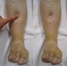
Characterized by noticeable fluid shifts within the lower extremities, ankles, and feet, commonly triggered by altered oncotic pressures or renal sodium retention channels.
An acute accumulation of fluid within the lungs’ alveolar spaces, indicating a medical emergency that leads to respiratory distress when lying flat.
Intracranial fluid accumulation capable of causing rapid pressure changes, often manifesting as neurological confusion or persistent headaches.
Swelling localized around the eyes or eye macula, frequently observed alongside heavy urinary protein loss in diabetic kidney conditions.
Primary Clinical Causes
Systemic fluid leakage into the tissue spaces is typically connected to several underlying medical concerns:
- • Advanced Kidney Pathologies: Severe protein leakage (Nephrotic Syndrome) or reduced baseline filtering efficiency.
- • Congestive Cardiac Insufficiency: Reduced pumping efficiency causing systemic venous pressures to back up.
- • Hepatic Cirrhosis: Impaired synthesis of serum albumin, lowering the blood’s natural holding capacity.
- • Microvascular Damage: Chronic vascular tracking secondary to prolonged hypertension or type-2 diabetes.
Identifying Diagnostic Indicators
When firm digital pressure applied to a swollen area leaves a persistent, visible depression or indentation that takes several seconds to smooth out, it points to freely movable interstitial fluid requiring formal medical review.
Associated Diagnostic Shifts
Keep a close watch for noticeably taut or reflective skin textures, unexplained rapid shifts on the scale, or sudden breathing changes when resting flat.
Strategic Clinical Interventions
Dr. Mukherjee focuses on diagnosing the primary physiological cause behind abnormal fluid shifts rather than temporarily masking visible swelling. Modern nephrological management protocols include:
Utilizing specialized diuretic therapies to encourage renal excretion of retained sodium and fluid volumes.
Addressing active proteinuria and managing serum albumin deficits to restore natural fluid holding capacities.
Designing personalized low-sodium dietary parameters to manage fluid retention without impacting lean nutrition.
Incorporating personalized graduated compression protocols and planned vascular elevation tracking.
Frequently Asked Clinical Questions
Pitting edema occurs when digital pressure leaves a persistent indentation in the skin, signaling movable fluid buildup due to systemic issues like kidney, heart, or liver conditions. Non-pitting edema doesn’t leave an indentation and typically points to localized lymphatic drainage blockages or thyroid dysfunction.
The kidneys maintain fluid and electrolyte balance. If filtering structures are damaged (as seen in Nephrotic Syndrome or Chronic Kidney Disease), they fail to excrete excess sodium or leak vital proteins like albumin into the urine. Low albumin levels reduce blood oncotic pressure, causing fluid to seep out into surrounding tissues as swelling.
Loop diuretics are highly effective for symptomatic relief by forcing the kidneys to flush out extra sodium and water. However, they are a management tool, not a permanent cure. Long-term management requires diagnosing and stabilizing the root cause, such as managing renal filtration errors or cardiovascular function.
Sodium acts like a sponge, pulling and retaining fluids inside the vascular and interstitial spaces. Even with medical diuretics, high salt intake keeps the fluid cycle going, overloading the kidneys and heart. A customized low-sodium diet stops the body from holding onto extra fluid right at the source.
Persistent Swelling Requires Specialist Assessment
Unexplained fluid retention is a key clinical indicator that merits a thorough diagnostic workup. Plan a formal assessment with our medical team today.
Doctor’s Desk
Dialysis Registry Desk
Sector 38, Gurugram