Kidney Biopsy Procedure
Advanced Percutaneous Kidney Biopsy Protocol
Precision Diagnostic Mapping by Prof. (Dr.) Debabrata Mukherjee, Senior Director of Nephrology & Renal Transplant, Gurugram.
Understanding the Renal Biopsy Process
A percutaneous kidney biopsy is considered the definitive clinical tool for diagnosing intricate parenchymal disorders. While non-invasive serum and urine assays provide functional clues, isolating a micro-fragment of renal tissue allows for comprehensive evaluation using light, immunofluorescence, and electron microscopy.
This procedure is approached with a commitment to patient comfort and structural precision. By integrating high-resolution, real-time ultrasound guidance with advanced target localization, we extract core samples efficiently while keeping internal risks to a minimum.
📌 Looking for the comprehensive step-by-step preparation checklist? Read our dedicated Kidney Biopsy Patient Guidelines Page for detailed dietary restrictions and medication pause-times.
When is a Histopathological Core Required?
When routine biomarkers leave diagnostic ambiguity, an expert tissue examination becomes essential to determine targeted therapy for several clinical presentations:
- ▪ Rapidly Progressive Glomerulonephritis (RPGN)
- ▪ Unexplained Cortical or Interstitial Lesions
- ▪ Acute Allograft (Transplant) Rejection Subtypes
- ▪ Refractory Nephrotic Syndrome in Adults
- ▪ Active Autoimmune Renal Damage (Lupus Nephritis)
- ▪ Persistent Isolated Hematuria and Proteinuria
The Biopsy Pathway: A Managed Timeline
Clinical Cleansing & Prep
Comprehensive evaluation of platelet function and clotting parameters (PT/INR). Complete dietary fasting is enforced for 6–8 hours prior, along with supervised pausing of all antiplatelet medications.
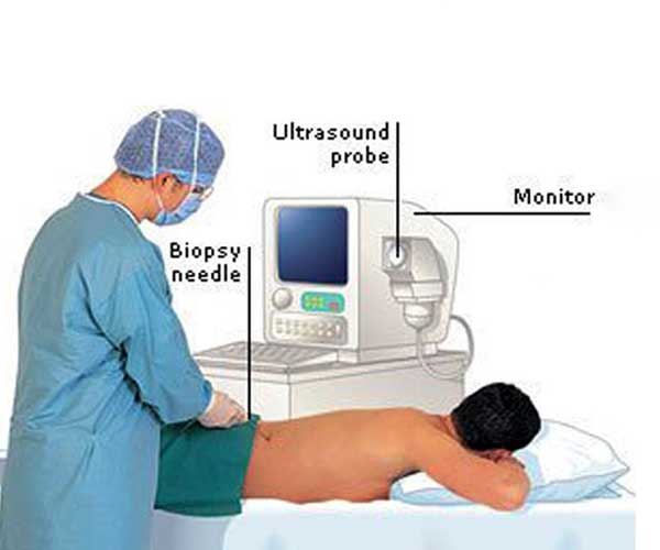
Ultrasound Core Extraction
Conducted under targeted local tissue anesthesia. Using automated, spring-loaded core biopsy guns guided by real-time ultrasound imaging, the cortical sample is retrieved within seconds.
Post-Procedure Vigilance
The patient rests in a flat position for 4–6 hours in our recovery ward. Vitals, urine color clear-times, and internal site stability are continuously checked before same-day or next-day discharge.
Evaluating Safety & Risk Controls
Modern percutaneous renal biopsies maintain an exceptional safety profile. Minor clinical events are handled with standard conservative steps:
- Localized muscular soreness near the needle path (resolves rapidly).
- Microscopic or transient hematuria (self-limiting within 24–48 hours).
- Extremely rare perinephric hematoma accumulation requiring extended monitoring.
Post-Procedure Stabilization
To support complete vascular sealing over the cortex, patients should observe these basic precautions during recovery:
- Restrict physical activities and demanding household exertion for 48 hours.
- Strictly avoid heavy weight-lifting or sudden core twisting (>5 kg) for 7–10 days.
- Maintain clean, dry site dressings for the initial 24 hours post-discharge.
- Follow specific hydration targets to support clear urinary flow.
Clinical FAQ: Understanding Your Kidney Biopsy
Q1: Is a kidney biopsy painful, and is it performed under general anesthesia?
Ans: No, a kidney biopsy is not performed under general anesthesia. It is done using local anesthesia, which numbs the skin and deeper tissue layers over the biopsy site. Patients remain awake and may feel a brief sensation of deep pressure when the automated needle captures the tissue sample, but the procedure itself is highly tolerable and virtually painless.
Q2: How long do I need to stop blood thinners before a renal biopsy?
Ans: Antiplatelet medications and anticoagulants (such as Aspirin, Clopidogrel, or Warfarin) must typically be discontinued 5 to 7 days prior to the procedure to minimize the risk of internal bleeding. This protocol must only be executed under the direct guidance of Prof. Dr. Mukherjee to ensure your primary cardiovascular condition remains stable.
Q3: What does blood in the urine after a kidney biopsy mean?
Ans: Mild or transient hematuria (pinkish-tinted urine) is a common and expected physiological event following a renal core extraction. It typically resolves naturally within 24 to 48 hours as the microscopic site clots. Continuous fluid intake is advised to prevent clot formation inside the bladder, and health metrics are tracked closely during your brief recovery stay.
Schedule Your Biopsy Evaluation
Achieve definitive histopathological clarity. Consult with our clinical team for optimized pre-procedural screenings and precise biopsy planning.
Clinical Daycare Liaison
Clinical Desk Communication: dirnephro@gmail.com
“`